An Expanding Frontier in Radiology and Cardiac Care
Back
Thoracic imaging is a critical field within radiology that is undergoing several significant changes due to ongoing advancements in artificial intelligence (AI).
Traditional chest X-rays and computed tomography scans –i.e., those methods most essential to diagnosing conditions like cardiomegaly, pleural effusion, and lung nodules-- are now being augmented by AI algorithms designed to improve accuracy, speed, and overall patient outcomes.
This technological maturation enhances the scope and reliability of AI tools in thoracic imaging.
Qure.ai is one of the key players in this evolving field. In October 2023, Qure received FDA clearance for its algorithm in cardiothoracic imaging, marking its 12th FDA-cleared algorithm.
FDA-Cleared AI Solutions and Their Impact
The FDA has cleared qXR-CTR, a deep learning-based algorithm developed by Qure.ai.
Qure specifically designed the algorithm to automate the measurement of the cardiothoracic ratio (CTR) on chest radiographs, a key indicator of cardiomegaly.
Not only does qXR-CTR deliver rapid results, but it also enhances diagnostic accuracy, thereby reducing the risk of heart failure. Moreover, it can screen for increased CTR and pleural effusion, two radiographic markers indicative of heart failure.
This advancement is monumental for the healthcare industry as it represents a tangible shift from speculative technology to clinically relevant applications. Dr. Tariq Ahmad of the Yale School of Medicine asserts that AI-driven solutions are increasingly helpful in narrowing the diagnostic gaps in cardiac care.
The effectiveness of AI algorithms in thoracic imaging is partly due to interdisciplinary collaborations among data scientists, radiologists, and cardiologists.
Such holistic expertise enriches the development and refinement of algorithms, ensuring they address real-world clinical challenges instead of acting in erroneous independence from them.
Critical Considerations for AI Integration
European Radiology outlines some of the critical steps necessary for the effective implementation of AI in radiology.
Researchers highlighted the need for a defined AI strategy and quality assurance, as is the necessity for comprehensive radiologist training on the limitations and functionalities of these AI tools.
It isn't a silver bullet; it is a tool that requires judicious application and continuous assessment.
As further highlighted in European Radiology, many tools have been developed with performances sometimes surpassing those of human radiologists, particularly in tasks like segmentation, detection, or classification.
AI in Low- and Middle-Income Countries
The Journal of the American College of Radiology provides valuable insights into deploying AI thoracic imaging in low- and middle-income countries (LMICs).
RAD-AID International uses a three-pronged strategy involving clinical education, infrastructure implementation, and phased AI introduction to deploy AI solutions in places like Guyana and Nigeria successfully.
However, the unique barriers in LMICs necessitate a tailored approach, underscoring the need for collaboration between local institutions and industry partners.
While technological advancements are often critiqued for their potential cost implications, AI algorithms such as qXR-CTR present an opportunity for cost efficiency.
By automating routine assessments, physicians can allocate more time to complex cases, optimizing resource allocation. This is particularly beneficial in low- and middle-income countries where medical resources may be scarce.
Unfurling the General Narrative of AI in Thoracic Imaging
Thoracic imaging is being reshaped by AI, forging a highly practical synergy of diagnostic precision and patient-centric care.
As AI matures from essential pattern recognition to sophisticated deep learning algorithms, it is carving out a niche in thoracic imaging, offering a lens of enhanced discernment in identifying and understanding thoracic anomalies.
Recent advancements underscore the potential of AI to transcend traditional diagnostic paradigms.
For instance, emerging techniques such as photon-counting computed tomography (CT) are being explored, potentially ushering in a new era of thoracic imaging with heightened sensitivity and specificity.
This, coupled with AI's algorithmic prowess, can significantly amplify the detection and characterization of thoracic pathologies, enabling a more nuanced understanding of disease processes.
Moreover, the transition of AI from a research endeavor to clinical practice is being catalyzed by rigorous validation processes, which pit algorithmic outputs against established medical guidelines and seasoned radiological expertise.
As noted in European Radiology, such a meticulous approach not only ensures the clinical relevance and safety of AI tools but also fosters trust among healthcare practitioners.
The journey of AI in thoracic imaging is a testimony to the power of interdisciplinary collaboration. The confluence of data scientists, radiologists, and cardiologists fuels the evolution of AI algorithms, ensuring they are fine-tuned to address real-world clinical challenges.
This collaborative ethos is at the heart of AI's growing footprint in thoracic imaging, promising a future where AI augments, not replaces, the human intellect in navigating the complexities of thoracic diseases.
As AI continues to make inroads into thoracic imaging, its implementation is challenging. The need for unbiased training and validation datasets, seamless workflow integration, and a clear understanding of legal and data protection requisites are critical for the successful deployment of AI.
Moreover, the onus is on the global healthcare community to ensure that the benefits of AI in thoracic imaging transcend geographical and economic boundaries, making quality healthcare accessible to all.
The advancement of AI in thoracic imaging is promising yet intricate. It is a journey from research to the bedside, from novelty to necessity, that could redefine the future of thoracic imaging and, by extension, cardiac care.
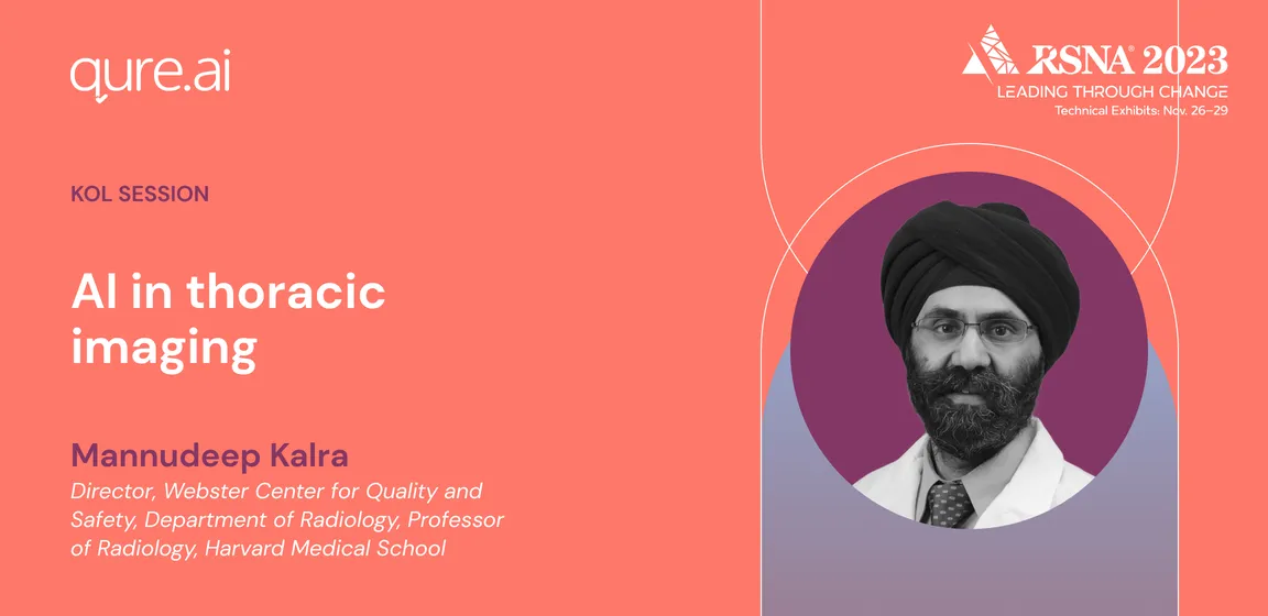
The assimilation of AI in thoracic imaging is a focal point of discussion in the upcoming RSNA chat. Dr. Mannudeep Kalra, a key speaker, brings a wealth of knowledge from his tenure at Massachusetts General Hospital and Harvard Medical School, where he's significantly invested in CT technology, radiation dose reduction, and AI research.
Register for the event here to explore the evolving frontier of AI in thoracic imaging.
