Musculoskeletal (MSK) radiology is rapidly evolving, with artificial intelligence (AI) playing an increasingly important role. AI-powered tools can improve the efficiency, accuracy, and accessibility of MSK imaging while providing new insights into diagnosing and treating MSK conditions.
Back
One of the most essential benefits of AI in MSK radiology is its ability to improve diagnostic accuracy.
AI is also making MSK imaging more accessible to patients. Radiologists can use AI to develop new imaging protocols that reduce scan time and radiation exposure. This is particularly important for patients who require frequent imaging, such as those with chronic MSK conditions.
The relationship between AI and MSK radiology is anchored in myriad applications that span image reconstruction, tissue segmentation, workflow augmentation, and disease detection.
The following outlines the significant avenues through which AI is sculpting the future of MSK radiology.
Augmenting Imaging Techniques
AI, particularly deep learning, has driven innovation in osteoarthritis, advancing image classification, lesion detection, cartilage segmentation, and beyond.
One of the significant drawbacks of MRI as a diagnostic tool has been the lengthy data acquisition time. However, AI has addressed this challenge head-on by accelerating MRI acquisition and reconstruction, thus reducing the long acquisition times that were previously a bottleneck.
This acceleration is crucial for large-scale longitudinal studies and emergency diagnostics where time is of the essence.
The 3D nature and soft-tissue contrast of MRI, which are indispensable for osteoarthritis research, are being significantly enhanced with the application of AI. AI facilitates the elucidation of disease pathogenesis and progression, making MRI an invaluable tool in osteoarthritis research. This enhancement in imaging quality provides a clearer view of the anatomical structures and the pathological changes occurring in osteoarthritis.
AI enables the automated detection of anatomically relevant health markers at the hip, knee, and ankle. This facilitates radiological monitoring of the progression of various bone diseases, such as knee osteoarthritis, thus providing a longitudinal view.
The application of AI, particularly machine learning and deep learning, is known to improve multiple stages of MRI, including acquisition, processing, and post-processing steps. This, in turn, minimizes the chances of diagnostic errors and enhances the overall efficacy and efficiency of osteoarthritis research.
Enriching Disease Understanding and Diagnosis
The automated models developed through AI have shown promise in staging knee osteoarthritis severity from radiographs, where the performance of these models has been comparable to that of musculoskeletal radiologists.
This automation ensures high diagnostic accuracy, critical for determining the appropriate treatment interventions.
Using AI and machine learning algorithms, researchers have identified early signs of osteoarthritis three years before diagnosis through MRI scans. This capability is essential for understanding the disease's developmental trajectory and identifying individuals who would likely benefit from early interventions.
AI improves the overall quality and efficiency of imaging, which is crucial for a more accurate diagnosis. It aids in assessing the appropriateness of imaging orders, thereby improving image quality, patient centricity, imaging efficiency, and, eventually, diagnostic accuracy.
Particularly in cases of advanced knee osteoarthritis requiring surgical intervention like Total Knee Arthroplasty (TKA), AI and machine learning modeling serve as indispensable tools in pre-surgical planning, patient selection, prediction of disease progression, and estimation of treatment outcomes. This is critical for tailoring the surgical approach to the individual needs and conditions of the patient, ensuring optimal post-surgical results.
Deep learning methods have been employed to improve the understanding of osteoarthritis.
AI transcends conventional diagnostic paradigms by facilitating a more comprehensive understanding of osteoarthritis by discovering novel image features indicative of short-term and long-term progression. This multi-faceted insight is crucial for a more patient-specific prognostic approach, ensuring the diagnostic deliberations are nuanced and rooted in a deeper understanding of the disease.
Enhancing Workflow and Clinical Operations
AI is identified as a pivotal tool that could significantly affect every step in the imaging value chain. The early integration of AI into radiology has already demonstrated its potential value, particularly in MSK imaging. The journey begins with placing the imaging requisition, traversing the entire imaging cycle, and ensuring well-oiled operational machinery.
The benefits of AI extend to workflow support, which is paramount for optimizing the efficiency and productivity of radiological practices. AI and deep learning offer multiple avenues of workflow support, including image reconstruction and transformation, tissue segmentation, and disease detection, which are intrinsic to MSK radiology.
Specifically, in spine imaging—a crucial part of MSK radiology—the incorporation of AI has led to significant advancements. Innovations rooted in AI and big data have considerably impacted musculoskeletal radiology, hinting at a future where a holistic approach could redefine every element of the radiology service value chain, from request to report.
From the broader radiology perspective, AI applications are being developed to span all steps of the imaging chain, from test ordering to report communication.
AI applications' traction across the radiology space underscores a promising horizon where workflow and delivery are redefined, thereby catalyzing a more efficient and streamlined operational framework in MSK radiology.
The role of qMSK
Amidst the evolving landscape of MSK radiology, the emergence of specialized AI tools drives nuanced advancements in diagnostic accuracy and operational efficiency.
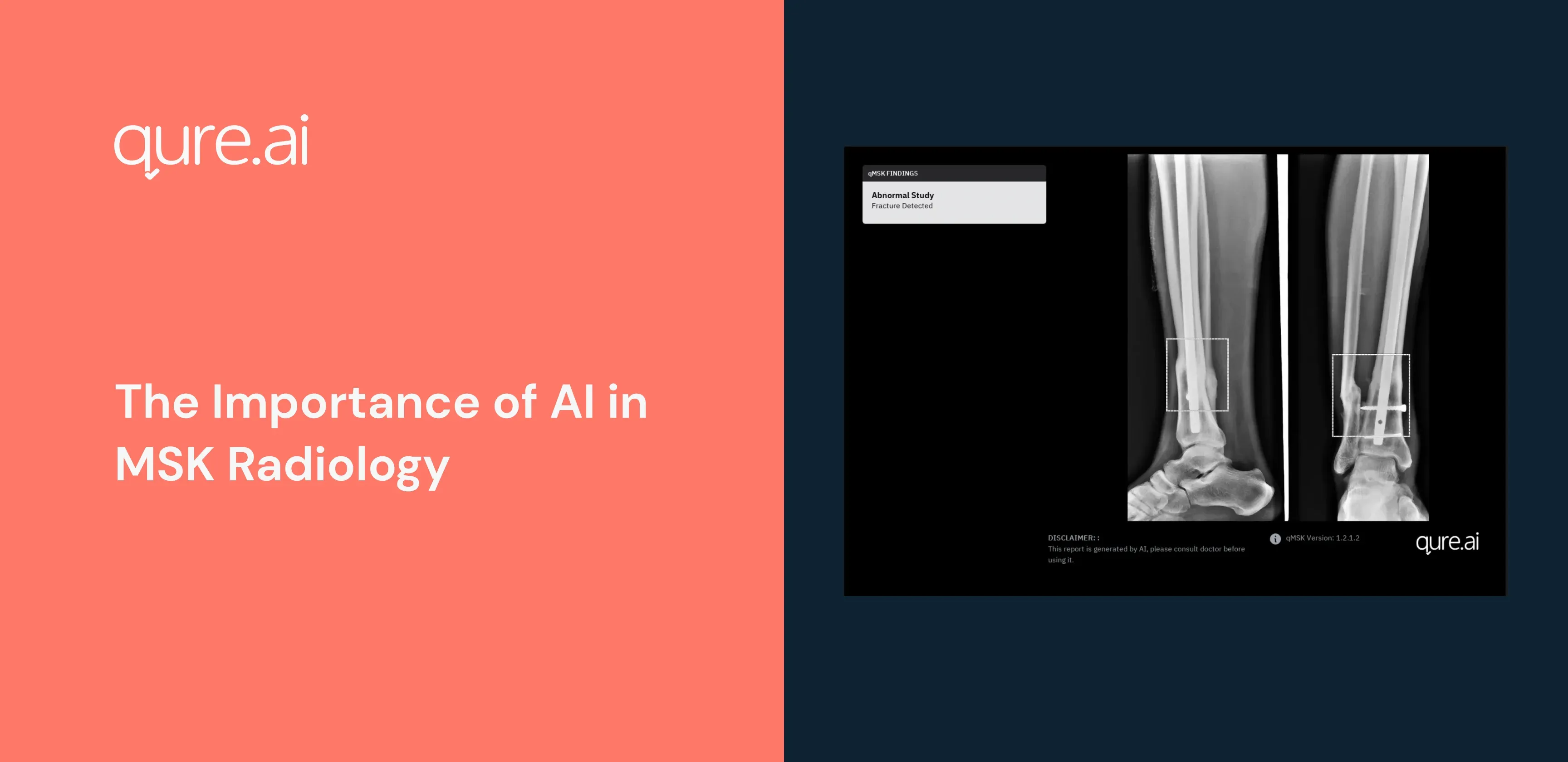
A notable example technologically in this trend is Qure.ai's qMSK software, engineered to augment the detection and localization of bone fractures and joint dislocations across multiple body regions on MSK X-rays, delivering insights in under a minute.
The comprehensive nature of qMSK extends to accommodating 15 anatomical views, thereby fostering a holistic understanding of potential musculoskeletal anomalies. This breadth of analysis is executed with a processing time of less than 20 seconds, testifying to the software's computational alacrity. Having been trained on over one million scans, qMSK exudes a sensitivity exceeding 0.9 in detecting signs of fractures, underscoring its diagnostic precision.
Moreover, qMSK is engineered to transcend conventional diagnostic thresholds by mitigating the 'satisfaction of search' bias - a phenomenon where the identification of one anomaly may overshadow or inhibit the detection of others.
By facilitating the recognition of multiple fractures, qMSK amplifies the diagnostic gaze, fostering a more comprehensive understanding of the patient's condition.
This capability is further enhanced by qMSK's proficiency in working across multiple views, thereby reducing diagnostic errors associated with overlooking subtle fractures.
The clinical journey of a patient in MSK radiology often necessitates a longitudinal examination of their condition, particularly in the context of fracture healing.
qMSK is tailored to track the progress in fracture healing, thus providing a dynamic diagnostic narrative pivotal for personalized patient care. Furthermore, the software significantly curtails the processing time to under 20 seconds per scan, translating to substantial time savings and augmented workflow efficiencywhen compounded over numerous cases.
In a domain where every second can be critical, qMSK's deployment flexibility resonates profoundly.
The software can be deployed on the cloud or on-premises, offering a versatile solution that aligns with the varied infrastructural and operational frameworks across different healthcare settings.
The articulation of qMSK's capabilities reflects a broader narrative of how AI bridges the technical and operational difficulties in MSK radiology by enhancing diagnostic acuity, streamlining the workflow, and catalyzing a more patient-centric approach.
Regulatory Recognition and Trajectory Forward
The institutional recognition of AI's efficacy is highlighted by the increasing number of AI products being cleared by the Food and Drug Administration (FDA) for utilization in clinical radiology, encompassing tasks such as quantification, identification, and even diagnosis.
This legitimizes AI's role in clinical radiology and hints at a trajectory of deeper integration shortly, with a broader spectrum of AI applications awaiting regulatory approval for clinical deployment.
Integrating AI in MSK radiology represents an enhancement and a fundamental transformation in diagnosing and treating musculoskeletal disorders.
This transformative alliance between AI and MSK radiology sets the stage for marked improvements in diagnostic precision, operational fluidity, and patient-centric care quality.