Key Using ultrasound AI to prevent cardiovascular diseases through early detection of atherosclerosis.
Back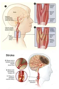
Key Highlights
- Cardiovascular diseases (CVD) cause ~32% of deaths; the leading cause of death globally
- qVH is the first known solution for AI-guided vascular ultrasound (carotid) that can boost disease prevention using point-of-care-ultrasound (POCUS) devices.
- With qVH, high-risk individuals can be screened by any clinician/ remote health worker at any convenient location
- Patients can be identified before symptoms appear, allowing for earlier disease management, improved patient outcomes, and reduced costs
Using AI to maximize the potential of POCUS for disease prevention
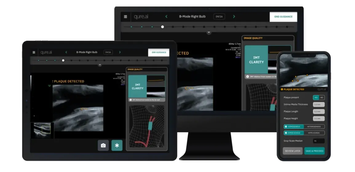
Qure.ai has developed an AI product for vascular health called qVH. It is the first known solution that guides clinicians during a carotid artery scan. Here’s how qVH’s AI works:
Probe Navigation Guidance: Detects probe location (CCA, ICA etc.) & orientation (long/short axis) and recommends best way to reach next step of protocol.
Plaque Detection & Characterization Guidance: Auto-detects abnormalities in a live scan (video) and quantifies it when a diagnostic quality image is available
Image Quality Guidance: Tracks image quality while scanning, recommends steps to maintain diagnostic quality & auto-captures images.
Device Setting Guidance: Detects common errors in ultrasound device settings in Pulse Wave (PW) mode and recommends changes for accurate PW velocity measurements. Thereby, preventing errors that could lead to misdiagnosis.
*qVH is not FDA approved/CE marked yet and is currently meant for investigational or research use only.
The need for qVH
~20% of strokes in adults are caused by the narrowing of the carotid artery (see image; bottom). Buildup of plaque (fatty deposits) in the arteries is the root cause for this narrowing; a condition that is known as atherosclerosis (see image; top).
Plaques can develop in different vessels leading to artery narrowing/clots and hence reduced blood flow to various parts of our body. This leads to critical events such as:
- Stroke (Carotid artery)
- Heart Attack (Coronary Artery)
- Renal Ischemia (Renal Artery), etc.
However, preventive measures to reduce disease burden have been limited due to:
- Lack of clear guidelines
- Logistical challenges like hospital visits for USG scan
- Depending on operator skills for accurate scanning & reporting using USG
- High cost of vascular USG
- Lack of sufficiently skilled clinicians to perform vascular USG.
Significant developments in the last decade
Price: USG machines have become cheaper (by ~80%), more portable (handheld & wireless) and easily accessible with the arrival of point-of-care ultrasounds (POCUS).
Preference: USG is gradually becoming the preferred modality for disease prevention.
- American Heart Association (AHA) recommends carotid artery duplex scanning in patients with high-risk features undergoing coronary artery bypass graft (CABG) surgery.
- European Society of Cardiology (ESC) recommends carotid duplex ultrasound for evaluating the extent and severity of carotid stenosis.
Proof: Real-world evidence suggests benefits in risk-stratification through carotid artery screening.
- Reduction in mortality rates (~10%), early disease diagnosis (~4.5 yrs), reduction in patient costs (by ~50%).
- Re-classification of low-risk patients to mid/high-risk based on the presence of carotid plaque. [Swiss AGLA]
Advantages: Govt. backed reimbursements for using advanced ultrasound technology.
- US Hospitals can claim NTAP reimbursement of ~$1868 per patient diagnosed using ultrasound guidance technology (Caption Guidance) for CVD prevention.
- American Medical Association (AMA) has introduced 2 new CPT codes for quantitative USG tissue characterization with ACR proposing an additional $82/scan.
What is holding back ultrasound-based CVD prevention programs?
POCUS devices have solved problems related to accessibility and affordability. But they have amplified issues related to operator skills since these ultra-portable POCUS devices can be used in any setting (remote areas, sports ground, battlefield etc.) by most clinicians. The existing issues are:
- Training is needed to perform the ultrasound scan as per the defined protocol.
- Training is needed to capture diagnostic quality images from a running video (cine loop).
- There are chances of misdiagnosis due to errors in:Performing ultrasound measurements manually. Eg: plaque length/area, PW ESV/PDV, Degree of Stenosis etc.Optimizing device settings manually. Ex: gate size/angle/position, box angle.Detecting & quantifying abnormalities (plaques, stenosis, etc.).
- Performing ultrasound measurements manually. Eg: plaque length/area, PW ESV/PDV, Degree of Stenosis etc.
- Optimizing device settings manually. Ex: gate size/angle/position, box angle.
- Detecting & quantifying abnormalities (plaques, stenosis, etc.).
- Inter-operator variability due to operator dependence for probe navigation, abnormality detection, image capture and for optimization of USG device settings.
qVH has been designed to address all of the issues outlined above. qVH validation has begun at 2 sites in India & Argentina and will be expanded to 8 sites across Asia, Europe and North America within the next 3 months. qVH can be used with all existing ultrasound machines. Our beta sites are using Cart and POCUS machines.